Instructions & Guidelines
Scroll down to access curated histopathology and tissue analytics resources including protocols, guidelines, and links to histology atlases.
Ordering & Sample Submission
Ordering via NUCORE
NUCORE Online Ordering InstructionsGo to NUCORE to place an order for MHPL Services
Sample Submission Instructions
Sample & Reagent Submission ChecklistMulti-Organ Submission Policy
A "multi-organ submission" refers to multiple tissue types and organs derived from a single experimental animal. Please read our MULTI-ORGAN SUBMISSION POLICY.
ISH/RNAscope Order Guidelines
MHPL Guidelines for In Situ Hybridization (ISH) and RNAscope Services
VALIDATING ORDER NUMBERS
Download step-by-step instructions to validate your order at NUCORE.
MHPL Service Rates
Download here the pricing for our services:
Effective 09/01/2025 until 08/31/2026
Organ Systems Development & Histology Atlases
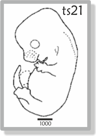
Staging Mouse Embryos
Mouse embryos can be staged according to a variety of criteria, the most general of which are those described by Theiler in "The House Mouse: Atlas of Mouse Development" (Springer-Verlag, New York, 1989).
Theiler Criteria for Staging Mouse Embryos
Atlas of Mouse Development
The following eHistology online resource provides free access to high-resolution colour images digitized from the original histological sections used by Kaufman for the Atlas of Mouse Development (1994).
![]()
Histology Atlas of the Mouse Placenta
The following paper provides an overview of histological transformations during the development of the mouse placenta which can be useful in identifying and describing changes in engineered, induced, and spontaneous disease models.
Histology Atlas of Mouse Urinary System Development
This review focuses on the histology of the mouse urinary system during embryonic development.
Histology Atlas of Mouse Heart Development
ATLAS OF MOUSE EMBRYONIC HEART DEVELOPMENTHistology of Mouse Lung Development
HISTOLOGY ATLAS OF MOUSE RESPIRATORY SYSTEM DEVELOPMENTHistology Atlas of Liver Development
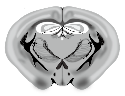
HISTOLOGY ATLAS OF MOUSE EMBRYONIC LIVER DEVELOPMENTBrain Atlases
The ALLEN BRAIN MAP is an excellent resource in neuroscience research and includes neuroanatomical atlases for mouse CNS, and human brain across multiple developmental timepoints.
Anatomy & Histology of the Pancreas
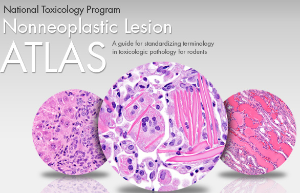
The PANCREAPEDIA is a useful resource for the anatomy and histology of the pancreas.Non-Neoplastic Lesion Atlas
The Non-Neoplastic Lesion Atlas hosted by the National Toxicology Program is a guide for standardizing toxicologic pathology in rodents. The atlas is subdivided into chapters covering a single organ.
Dissection, Grossing & Fixation
Sampling of Rodent Organs for Histology
The following papers summarize suggested guidelines for preparing various organs in tissues from rodents for histological analysis.
Tissue Collection for Systematic Mouse Phenotyping
Respiratory, Reproductive and Thymic Organs
Neuronal, Urogenital Tract, Cardiovascular, and Immune Organs
Intestinal Swiss Rolls
Download the detailed JOVE protocol for preparing intestinal Swiss rolls.Watch the video to prepare intestinal Swiss rolls for histology
Fixation of Lungs for Histology
Transcardial Perfusion-Fixation
This method is the preferred fixation strategy for CNS tissues and also to flush out blood from organs.
Glyoxal Fixation
Glyoxal can replace formaldehyde-based fixatives (10% NBF or 4% PFA) as a fast-acting cross-linker that also preserves protein and nucleic acid antigenicity. Glyoxal-fixation typically results in brighter microscopy images.
Lipid Fixation with Chromic Acid
Chromic acid can fix lipids in tissues prior to creation of FFPE blocks. This is a convenient alternative to cryosections for evaluation of lipid accumulation.
Fixation of Ocular & Testicular Specimens
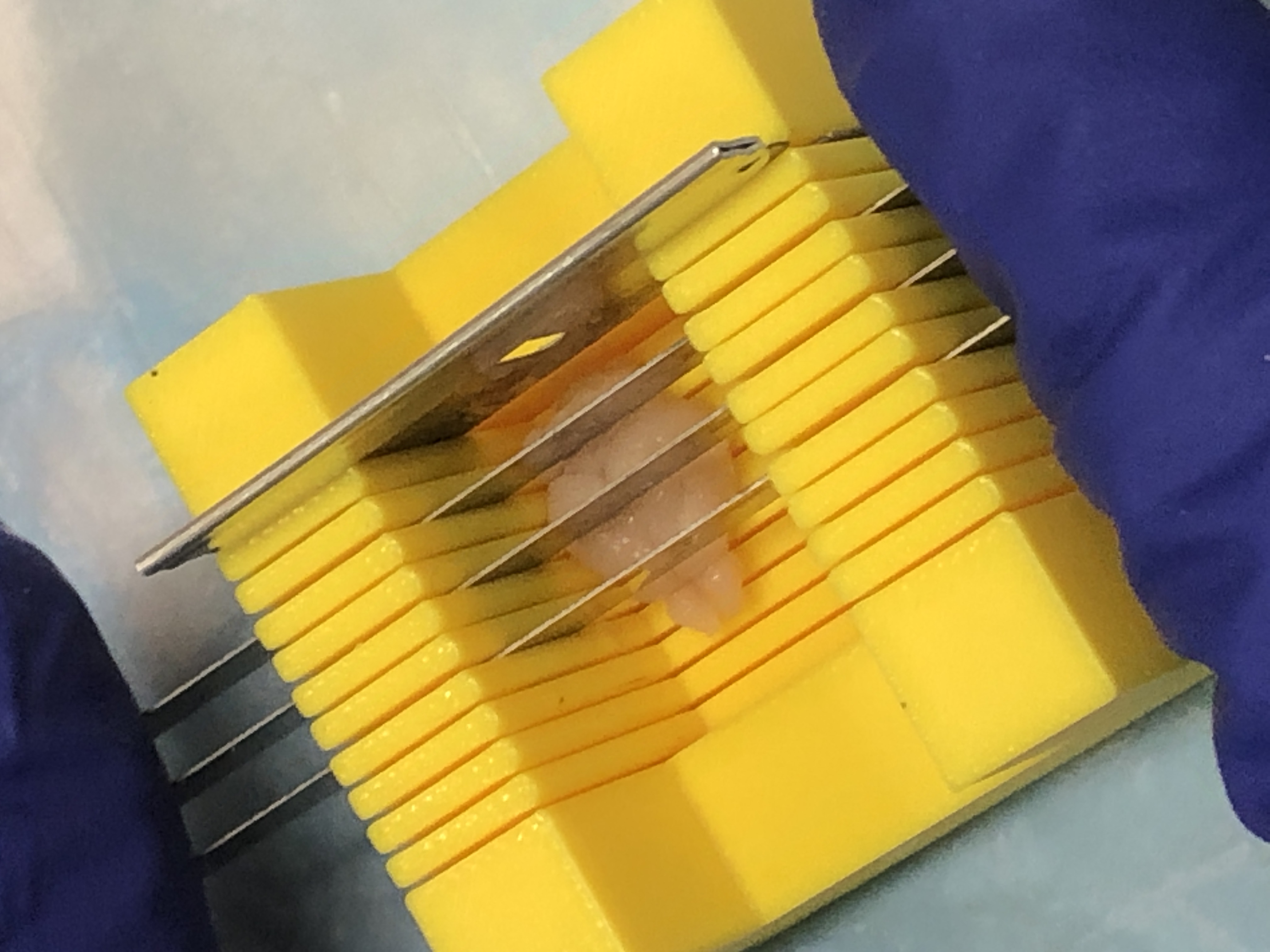
Heart Tissue Processing for Infarct Analysis
This protocol describes the use of a tissue matrix to sample thick sections of the heart for a systematic survey of myocardial infarct.
Decalcification Protocols
Decalcification with Immunocal
This method is a formic acid-method using IMMUNOCAL for decalcifying mineralized tissues to make them suitable for microtomy and subsequent histology and immunostainings.
Decalcification with EDTA
This method for decalcification utilizes a buffered EDTA-sucrose solution. This method is suitable for spatial transcriptomics to maintain the integrity of RNA in the tissues.
Paraffin Microtomy Resources
Rotary Microtome Self-Service Guidelines
MHPL Rotary Microtome Self-Service GuidelinesMicrom HM325 Microtome Manual
Microm HM325 Microtome User ManualParaffin Microtomy Tips & Additional Resources
Download the following useful resource on paraffin microtomy from Leica:
Microtomy & Paraffin Section Preparation
Cryosectioning Resources
Cryostat User Guidelines
Dakewe CT520 Cryostat Manual & Instructional Video
Dakewe CT520 User Manual (Full)
Cryosectioning Tutorial
Cryostat TutorialVibratome Resources
Vibratome Self-Service Policies
Vibratome User GuidelinesLeica VT1200S User Manual
Leica VT1200S ManualVibratome Sectioning of Rodent Brain Slices
Sectioning of Thick Liver Slices
Liver Sectioning using the VibratomeOptimization of Vibratome Sectioning
Vibratome Troubleshooting for Better SectionsPreparation of Small Specimens for Histology
Histogel Embedding
Histogel Embedding ProtocolAlginate Embedding
Alginate Embedding ProtocolPrepare Slides of Single Cell Suspensions
The Cytospin method prepares single cell suspension smears on glass slides by centrifugation suitable for downstream histology, immunostaining, or nucleic acid in situ hybridization.
In Situ Hybridization Resources
Multiplex Fluorescent RNAscope
Protease-Free Multiplex RNAscope ProtocolmiRNAscope & RNAscope Plus Assays
miRNAscope HD-Red Assay (Chromogenic/Fluorescent)
RNAscope Plus (RNAscope and small RNA co-detections-Fluorescent)
Tissue Clearing Resources
X-CLARITY Tissue Clearing
X-CLARITY Tissue Clearing ManualX-CLARITY Tissue Clearing Protocol
X-CLARITY Electrophoretic Tissue Clearing ProtocolSelection Guides for Reagents, Antibodies and Control Tissues
Selection of Antibodies
CITEAB is a useful website summarizing applications and published citations for commercially available antibodies.
![]()
Selecting Positive Control Tissues
Here are useful resources for identifying positive control tissues for immunolabelings and in situ hybridization:
FLUORESCENCE SPECTRA VIEWER
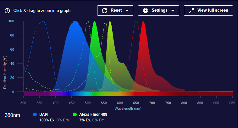
Image Analysis
Fiji / ImageJ
FIJI/ImageJ is a widely used, free, and open-source software platform for scientific image analysis.
![]()
QuPATH
QuPATH is an open-source, cross-platform software application primarily used for bioimage analysis, particularly within the context of digital pathology and whole slide image analysis.
![]()
CellProfiler
CellProfiler is a free, open-source image analysis software designed for biologists to automatically identify, quantify, and analyze biological entities in images. It allows users to create custom pipelines using various image processing and object analysis modules, facilitating reproducible and quantitative analysis of cell and subcellular features.
![]()
RNAscope Data Analysis
This ACD web resource provides a wealth of information on how to analyze RNAscope data in chromogenic singleplex/duplex to multiplex fluorescent scenarios.
CellProfiler Analysis of RNAscope & BASEscope Data
This ACD web resource provides instructions on how to analyze RNAscope or BASEscope results using CellProfiler.
PIXIMI
Piximi is an open-source, browser-based platform for interactive bioimage analysis. Designed for accessibility and flexibility, Piximi enables users to perform advanced image processing tasks without requiring any installation or deep machine learning expertise. Built with researchers and biologists in mind, it provides a user-friendly interface for annotation, segmentation, classification, and quantitative measurement of microscopy images.Miscellaneous
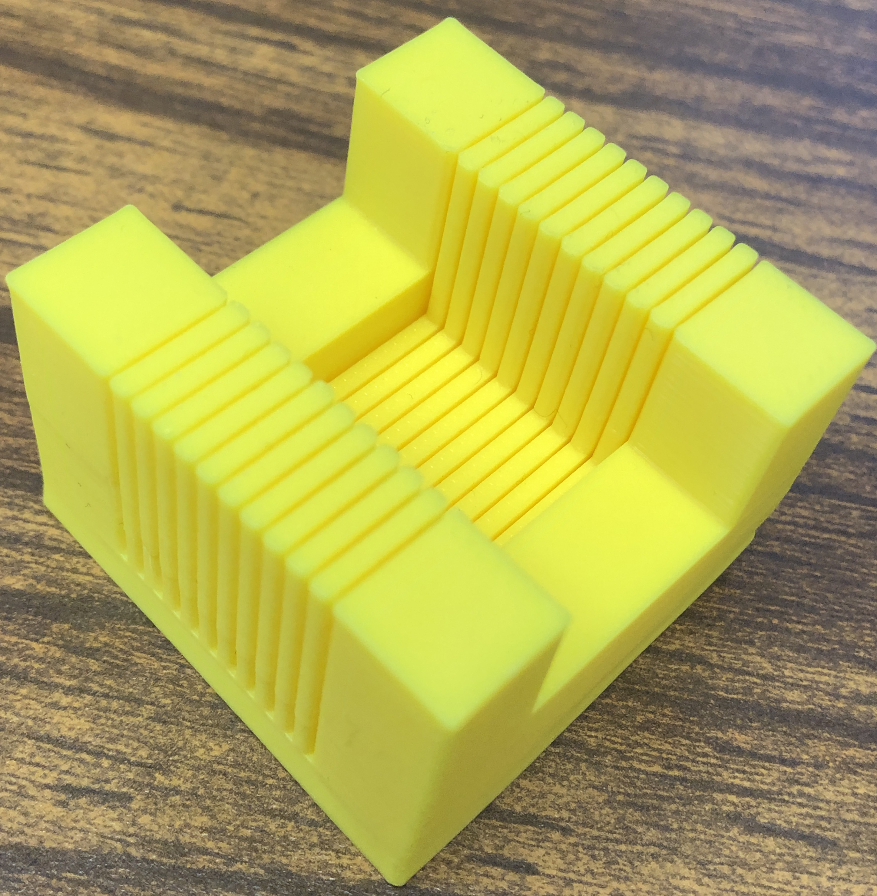
3D Printing Brain Matrices
This protocol provides instructions on how to 3D print mouse brain slicing matrices.
3D Printing Services are available at:
MakerLab at the Mudd Library in the Evanston campus.
CAM/NIC at Searle Bldg. in the Chicago campus.
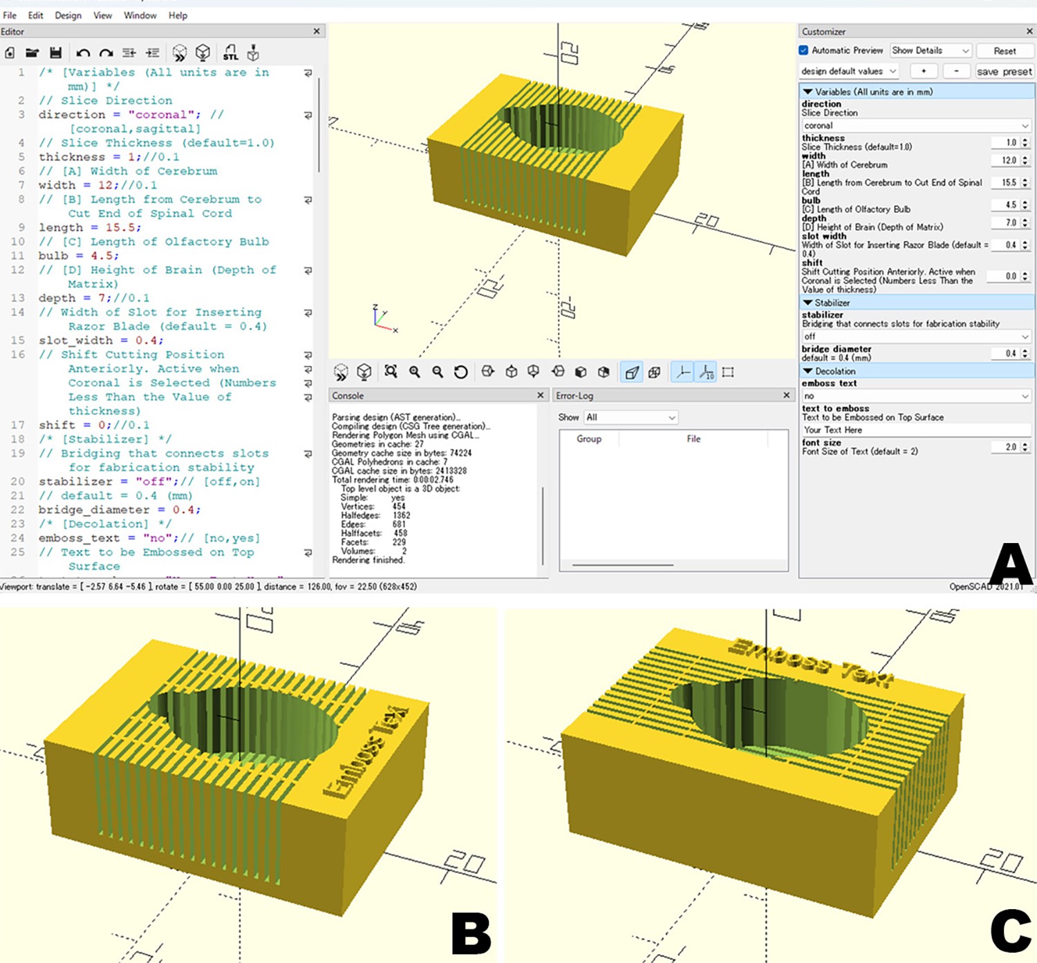
3D Printing of Tissue Sectioning Matrix
Tissue Matrix 3D Printing Template
3D Printing Services are available at:
MakerLab at the Mudd Library in the Evanston campus.
CAM/NIC at Searle Bldg. in the Chicago campus.
Thick Slicing of Brain & Heart Using a Tissue Matrix
This instructional video shows how to systematically slice thick sections of brain and heart tissues using a tissue matrix.